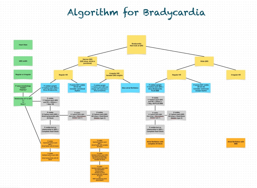
- Overview of Dubin’s Method for Reading EKG’s
- 1. RATE (pp. 65-96)
- 2. RHYTHM (pp. 97-202)
- 3. AXIS (pp. 203-242)
- 4. HYPERTROPHY (pp. 243-258)
- 5. INFARCTION (pp. 259-308)
- Further discussion
- Rate (pp. 65-96)
- Rhythm (pp. 97-111)
- Rhythm – continued (pp. 112-172)
- Rhythm (Heart Blocks) (pp. 173-202)
- Axis (pp. 203-242)
- Hypertrophy (pp. 243-258)
- Infarction (pp. 259-308)
- Infarction Location/Coronary Vessel Involvement
- Miscellaneous (pp. 309-328)
- Practical Tips
Overview of Dubin’s Method for Reading EKG’s
1. RATE (pp. 65-96)
Estimate using “300, 150, 100…”
Bradycardia: rate = cycles/6 sec. strip × 10
2. RHYTHM (pp. 97-202)
Identify basic rhythm, scan for prematurity, pauses, irregularity, abnormal waves. Check for:
- P before each QRS, QRS after each P
- If Axis Deviation, rule out Hemiblock.
3. AXIS (pp. 203-242)
QRS above or below baseline for Axis Quadrant (Normal vs. R. or L. Axis Deviation).
Find isoelectric QRS in a limb lead for Axis in degrees using the “Axis in Degrees” chart.
Axis rotation (horizontal plane): find “transitional” (isoelectric) QRS.
4. HYPERTROPHY (pp. 243-258)
Check P wave (atrial hypertrophy).
Check R wave in V₁ (Right Ventricular Hypertrophy).
Check S wave depth in V₁ and R wave height in V (Left Ventricular Hypertrophy).
5. INFARCTION (pp. 259-308)
Scan leads for:
Q waves
Inverted T waves
ST segment elevation/depression
Find location of pathology and identify the occluded coronary artery.
Further discussion
Rate (pp. 65-96)
Determine rate by observation using the triplet method (300-50).
Fine division/rate association reference chart provided.
- Bradycardia: cycles/6 second strip × 10 = rate
- Sinus Rhythm: SA Node origin; normal rate 60-100/min.
100/min = Sinus Tachycardia
<60/min = Sinus Bradycardia
Determine independent (atrial/ventricular) rates if co-existing rhythms are present
- Dissociated Rhythms: sinus or atrial rhythm can co-exist with an independent rhythm from a focus of a lower level
- Irregular Rhythms: note average ventricular rate (QRS’s/6-sec strip x 10)
Rhythm (pp. 97-111)
Identify basic rhythm, then scan tracing for pauses, premature beats, irregularity, and abnormal waves.
Always check: P before each QRS, QRS after each P; PR intervals, QRS interval; QRS vector shift outside normal range
- Sinus Arrhythmia: Irregular, varies with respiration, all P waves identical
- Wandering Pacemaker: Irregular, P waves change shape, rate < 100/minute
- Multifocal Atrial Tachycardia: Rate >100/min; otherwise similar to Wandering Pacemaker
- Atrial Fibrillation: Irregular ventricular rhythm, no P waves (erratic atrial spikes)
Rhythm – continued (pp. 112-172)
- Escape (pp. 112-121) – The heart’s response to a pause in pacing.
An unhealthy Sinus (SA) Node fails to emit a pacing stimulus (“Sinus Block”).
A sick Sinus (SA) Node may cease pacing (“Sinus Arrest”).
Escape Beats can be: Atrial, Junctional, or Ventricular.
- Premature Beats (pp. 122-145) – From an irritable automaticity focus.
Premature Beats can be: Atrial, Junctional, or Ventricular.
- Tachyarrhythmias (pp. 146-172)
Paroxysmal – rate: 150-250/min.
Flutter – rate 250-350/min
Fibrillation – rate 350-450/min
Rhythm (Heart Blocks) (pp. 173-202)
- Sinus (SA) Block: An unhealthy sinus node misses one or more cycles; The SA Node usually resumes pacing, but the pause may evoke an “escape” response from an automaticity focus.
- AV Block: Blocks that delay or prevent atrial impulses from reaching the ventricles.
1st AV Block: Prolonged PR interval
2nd AV Block: Some P waves without QRS response
Wenckebach
Mobitz
3rd AV Block: No P wave produces a QRS response
Axis (pp. 203-242)
- General determination – is QRS (+) or (-) in leads I and AVF
- First determine axis quadrant
if the QRS is positive in I and AVF = Normal
Axis in Degrees
After locating the Axis Quadrant, find the limb lead where QRS is most isoelectric
- Axis rotation (left/right) in the Horizontal Plane:
Find transitional (isoelectric) QRS in a chest lead
Hypertrophy (pp. 243-258)
Right Atrial Hypertrophy: Large, diphasic P wave with tall initial component
Left Atrial Hypertrophy: Large, diphasic P wave with wide terminal component
Right Ventricular Hypertrophy
Left Ventricular Hypertrophy
Infarction (pp. 259-308)
- Q wave = Necrosis (significant Q’s only)
- ST segment elevation = (acute) Injury (also Depression)
- T wave inversion = Ischemia
Always obtain patient’s previous EKG’s for comparison!
Infarction Location/Coronary Vessel Involvement
Know Coronary Artery Anatomy.
Posterior
Lateral
Inferior
Anterior
Miscellaneous (pp. 309-328)
Pulmonary Embolism
Artificial Pacemakers
Electrolytes:
Potassium
Calcium
Digitalis
Quinidine
Practical Tips
Dubin’s Quickie Conversion: Patient’s weight in kg. = Half of patient’s wt. (in lb.) minus 1/10 of that value.
Modified Leads for Cardiac Monitoring.