Managing asthma
Continue readingCOPD initial management
Here the initial GOLD algorithm
Continue readingChronic fatigue workup
Start with a focused history, exam, and a limited, high-yield lab panel to rule out common, reversible causes; then use symptom-directed testing.
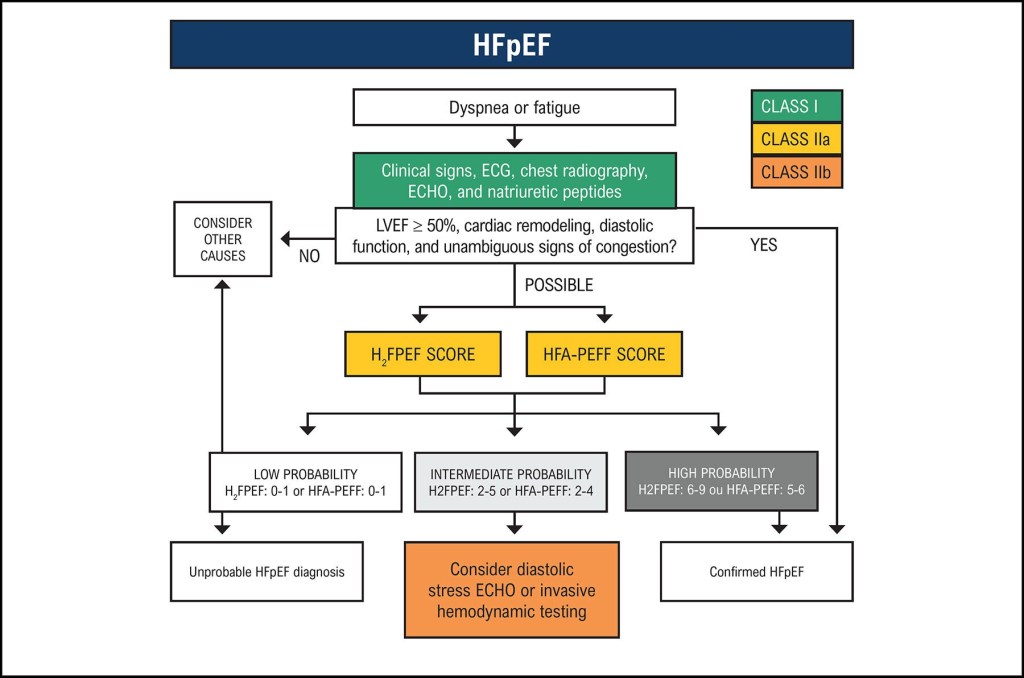
Continue readingHFpEF: overview of management of heart failure with preserved ejection fraction
Suggested reading:
Up-to-date
Overview:
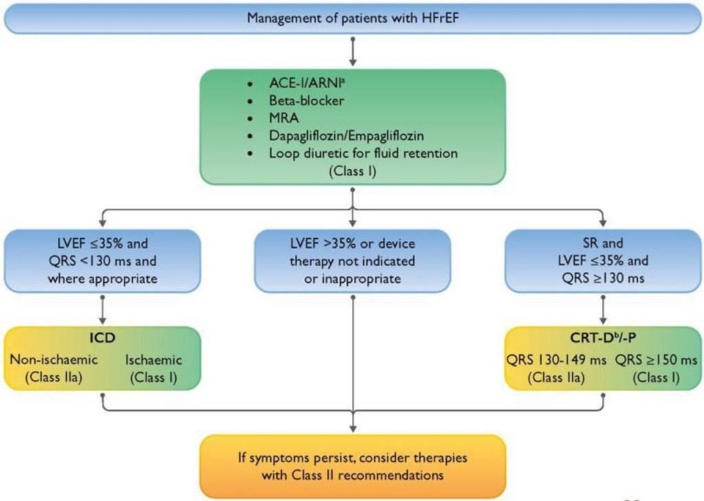
HFrEF: overview of management of heart failure with reduced ejection fraction
Suggested reading:
Up-to-date
Overview:
EKG: Tachycardia
Steps in evaluation
Step one: verify tachycardia and hemodynamic stability. If unstable refer to ACLS algorithms please. 
Step two: determine whether the QRS is narrow complex or wide complex
Step three: if narrow complex determine whether irregular or regular rhythm
Step four: if wide complex tachycardia, determine whether monomorphic or polymorphic
Algorithm for the initial ECG review and differential diagnosis of tachycardia

ECG: electrocardiogram; AVNRT: atrioventricular nodal reentrant tachycardia; AVRT: atrioventricular reciprocating (bypass-tract mediated) tachycardia; AT: atrial tachycardia; SANRT: sinoatrial nodal reentrant tachycardia; AF: atrial fibrillation; AV: atrioventricular; VT: ventricular tachycardia; SVT: supraventricular tachycardia; WPW: Wolff-Parkinson-White.
* A narrow QRS complex is <120 milliseconds in duration, whereas a wide QRS complex is ≥120 milliseconds in duration.
¶ Refer to UpToDate topic reviews for additional details on specific ECG findings and management of individual arrhythmias.
Δ Monomorphic VT accounts for 80% of wide QRS complex tachycardias; refer to UpToDate topic on diagnosis of wide QRS complex tachycardias for additional information on discriminating VT from SVT.
Algorithm for the evaluation of narrow QRS complex tachycardia in stable patients

How to read EKGs
- Overview of Dubin’s Method for Reading EKG’s
- Further discussion
- Miscellaneous (pp. 309-328)
- Practical Tips
Overview of Dubin’s Method for Reading EKG’s
1. RATE (pp. 65-96)
Estimate using “300, 150, 100…”
Bradycardia: rate = cycles/6 sec. strip × 10
2. RHYTHM (pp. 97-202)
Identify basic rhythm, scan for prematurity, pauses, irregularity, abnormal waves. Check for:
- P before each QRS, QRS after each P
- PR intervals (AV Blocks)
- QRS interval (BBB)
- If Axis Deviation, rule out Hemiblock.
3. AXIS (pp. 203-242)
QRS above or below baseline for Axis Quadrant (Normal vs. R. or L. Axis Deviation).
Find isoelectric QRS in a limb lead for Axis in degrees using the “Axis in Degrees” chart.
Axis rotation (horizontal plane): find “transitional” (isoelectric) QRS.
4. HYPERTROPHY (pp. 243-258)
Check P wave (atrial hypertrophy).
Check R wave in V₁ (Right Ventricular Hypertrophy).
Check S wave depth in V₁ and R wave height in V (Left Ventricular Hypertrophy).
5. INFARCTION (pp. 259-308)
Scan leads for:
Q waves
Inverted T waves
ST segment elevation/depression
Find location of pathology and identify the occluded coronary artery.
Further discussion
Rate (pp. 65-96)
Determine rate by observation using the triplet method (300-50).
Fine division/rate association reference chart provided.
- Bradycardia: cycles/6 second strip × 10 = rate
- Sinus Rhythm: SA Node origin; normal rate 60-100/min.
100/min = Sinus Tachycardia
<60/min = Sinus Bradycardia
Determine independent (atrial/ventricular) rates if co-existing rhythms are present
- Dissociated Rhythms: sinus or atrial rhythm can co-exist with an independent rhythm from a focus of a lower level
- Irregular Rhythms: note average ventricular rate (QRS’s/6-sec strip x 10)
Rhythm (pp. 97-111)
Identify basic rhythm, then scan tracing for pauses, premature beats, irregularity, and abnormal waves.
Always check: P before each QRS, QRS after each P; PR intervals, QRS interval; QRS vector shift outside normal range
- Irregular Rhythms:
- Sinus Arrhythmia: Irregular, varies with respiration, all P waves identical
- Wandering Pacemaker: Irregular, P waves change shape, rate < 100/minute
- Multifocal Atrial Tachycardia: Rate >100/min; otherwise similar to Wandering Pacemaker
- Atrial Fibrillation: Irregular ventricular rhythm, no P waves (erratic atrial spikes)
Rhythm – continued (pp. 112-172)
- Escape (pp. 112-121) – The heart’s response to a pause in pacing.
An unhealthy Sinus (SA) Node fails to emit a pacing stimulus (“Sinus Block”).
A sick Sinus (SA) Node may cease pacing (“Sinus Arrest”).
Escape Beats can be: Atrial, Junctional, or Ventricular.
- Premature Beats (pp. 122-145) – From an irritable automaticity focus.
Premature Beats can be: Atrial, Junctional, or Ventricular.
- Tachyarrhythmias (pp. 146-172)
Paroxysmal – rate: 150-250/min.
Flutter – rate 250-350/min
Fibrillation – rate 350-450/min
Rhythm (Heart Blocks) (pp. 173-202)
- Sinus (SA) Block: An unhealthy sinus node misses one or more cycles; The SA Node usually resumes pacing, but the pause may evoke an “escape” response from an automaticity focus.
- AV Block: Blocks that delay or prevent atrial impulses from reaching the ventricles.
1st AV Block: Prolonged PR interval
2nd AV Block: Some P waves without QRS response
Wenckebach
Mobitz
3rd AV Block: No P wave produces a QRS response
Axis (pp. 203-242)
- General determination – is QRS (+) or (-) in leads I and AVF
- First determine axis quadrant
if the QRS is positive in I and AVF = Normal
Axis in Degrees
After locating the Axis Quadrant, find the limb lead where QRS is most isoelectric
- Axis rotation (left/right) in the Horizontal Plane:
Find transitional (isoelectric) QRS in a chest lead
Hypertrophy (pp. 243-258)
- Atrial Hypertrophy
Right Atrial Hypertrophy: Large, diphasic P wave with tall initial component
Left Atrial Hypertrophy: Large, diphasic P wave with wide terminal component
- Ventricular Hypertrophy
Right Ventricular Hypertrophy
Left Ventricular Hypertrophy
Infarction (pp. 259-308)
- Q wave = Necrosis (significant Q’s only)
- ST segment elevation = (acute) Injury (also Depression)
- T wave inversion = Ischemia
Always obtain patient’s previous EKG’s for comparison!
Infarction Location/Coronary Vessel Involvement
Know Coronary Artery Anatomy.
Posterior
Lateral
Inferior
Anterior
Miscellaneous (pp. 309-328)
Pulmonary Embolism
Artificial Pacemakers
Electrolytes:
Potassium
Calcium
Digitalis
Quinidine
Practical Tips
Dubin’s Quickie Conversion: Patient’s weight in kg. = Half of patient’s wt. (in lb.) minus 1/10 of that value.
Modified Leads for Cardiac Monitoring.
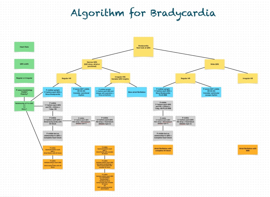
EKG: Bradycardia
Here is the algorithm for bradycardia
Insulin regimens
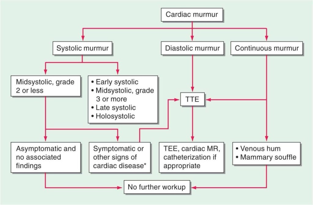
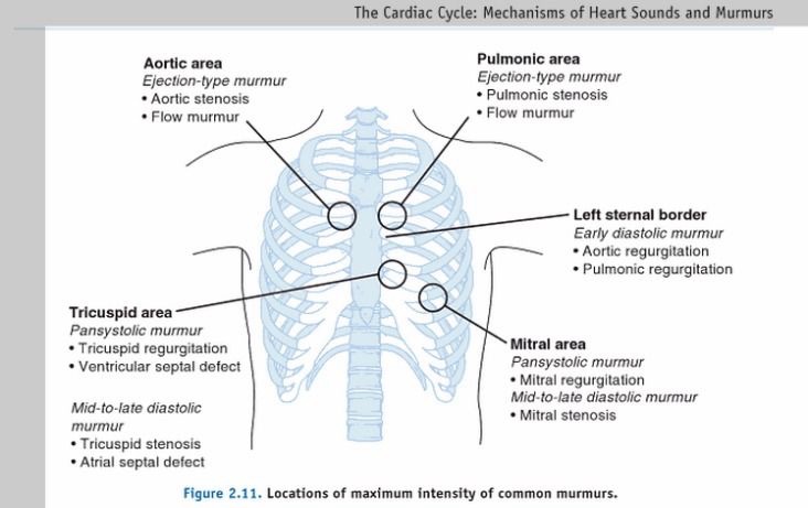
Heart murmurs
Murmur location
Systolic vs diastolic and Murmur description

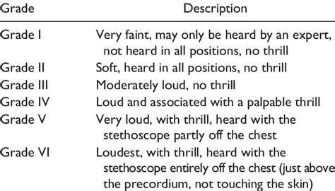
Murmur grade (Levine’s grading)
How to approach heart murmurs