
How to approach LBP.
It consists of inspection, Palpation, special tests, and Neurological exam.
Inspection
The first part of the low back exam starts with inspection. First, note the contour of the spine. Appreciate the normal posterior curvature of the upper spine (kyphosis) and the normal anterior curvature of the lower spine (lordosis). Lack of lumbar lordosis (i.e. a flat lower spine) is often associated with low back pain.
Palpation
For the second part, palpation, we generally focus on two areas:
1) The center of the back or the spinal region. Pain here suggests pain from the vertebra
2) Just lateral to the center or para-spinal regions. Pain here suggests pain from a muscle strain of the paraspinal muscles.
Special Tests
Straight Leg Test
 The most common provocative test is the straight leg test. To conduct this test, have the patient lay supine and passively elevate the fully extended leg of the affected side to 30-60 degrees. You should need to extend the leg more than 60 degrees. A positive test will elicit pain in the region where the patient was complaining of pain in the back, often radiating down the leg.
You can often elicit pain in the affected side by lifting the leg on the other side if the nerve irritation is severe enough.
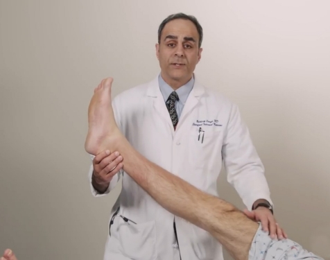
Straight Leg Test Variant
 Another variant of the straight leg test involves lower the leg to around 30 degrees and flexing the foot as depicted in the image. As in the straight leg test, a positive test involves pain in the lower back, often radiating down the leg.
Tripod Sign
 The tripod sign is a provocative test that is conducted while the patient is in the seated position. By elevating one of the legs, a positive sign will elicit pain in the back (again often radiating down the leg) and should be accompanied by the patient’s natural tendency to decrease the pain by leaning back and resting both arms on the table to support him or herself, thus the creating a tripod.
Note: this is a good sign to use with patient’s suspected of malingering if they complain of pain. Failure to lean back and rest both arms on the table may suggest the pain is not present or not related to irritation of the nerve roots.
Femoral Stretch Test (L2-4)
 The straight leg and tripod signs are more sensitive to pain in the L5 & S1 regions. If you suspect pain coming from the L2-4 region (which is less common), you can test for it with the femoral stretch test.
This test is done by having your patient lie prone on their stomach. Next, flex the leg at the knee while holding the base of the left under the knee. Next, simply lift the whole leg up. A positive test suggests pain in the L2-4 region if they complain of pain in the anterior thigh while the leg is lifted up.
The Neurologic Exam
The neurological exam consists:
1) Motor Exam
2) Sensory Exam
3) Reflex Exam
Of note, the major nerve roots to examine include L4, L5, and S1 as they are the most commonly affected. Therefore, we will focus on these three roots as well for each neurological exam.
The Motor exam
L4 Motor Exam
 To test L4 strength, have the patient slightly bend the knee and kick out as you keep pressure against the leg. Be sure to compare both sides to see if one side has weakness relative to the other.
L5 Motor Exam
 Dorsiflexion of the big toes.
To test L5 strength, hold pressure over the large toes and ask the patient to dorsiflex the big toes and foot towards up. Compare both sides for relative weakness.
S1 Motor Exam
To test S1 strength, hold pressure under both feet and ask the patient to plantarflex the foot down. Compare both sides for relative weakness.


Test for L5 weakness with walking on heels in normal patients. If one foot is unable to lift toes off the ground, could suggest L5 weakness on that side.

Test for S1 weakness with walking on toes in normal patients. If one foot is unable to lift heal off the ground, could suggest S1 weakness on that side.
The Sensory exam
L4 Sensory Exam
Focus on the anterior/lateral aspect of the thigh.

L5 Sensory Exam
Focus on the space on the dorsal side between the first and second toe.

S1 Sensory Exam
Focus on the lateral aspect of the foot.

The Reflex exam
The last part of the neurological assessment is the reflex exam. Again we look at L4, L5 & S1. Pay attention to differences on either side.
L4 Reflex Exam
L4 is tested by the patellar reflex.

L5 Reflex Exam
L5 is tested by the medial hamstring reflex.

S1 Reflex Exam
S1 is tested by the Achilles reflex.

